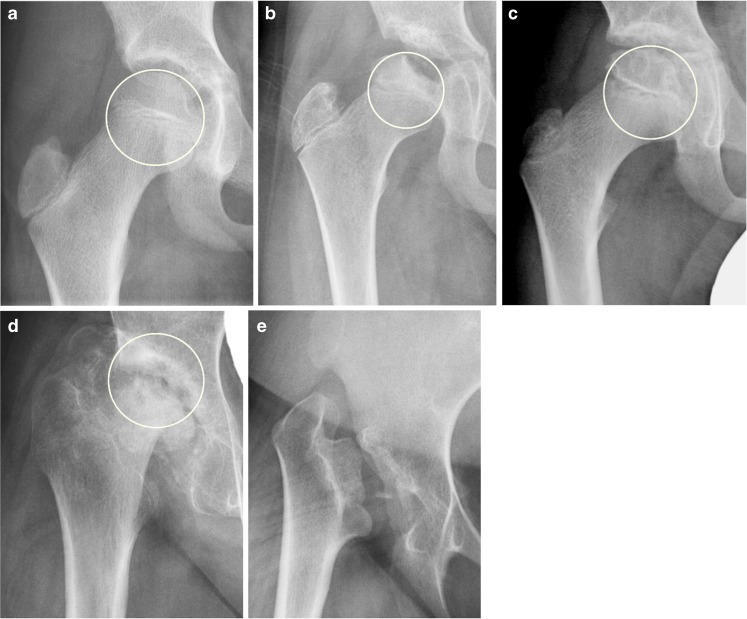
Fig. 2.
Femoral head flattening of differing severities in children and a young adult with juvenile idiopathic arthritis, as shown on an anteroposterior pelvic radiograph measured by the Mose template for reference. The Mose template in (a–d) is a circle drawn to represent where the femoral head should be located, and the degree of flattening is judged using a score of 0–4 according to 25% incremental losses in head height. a Radiograph in a 9-year-old boy shows normal femoral head without any loss of height (score = 0). b Radiograph in a 10-year-old boy shows mild loss of femoral head height of <25% (score 1). c Radiograph in a 9-year-old boy shows moderate loss of femoral head height of 26–50% (score 2). d Radiograph in a 13-year-old boy shows marked loss of femoral head height of 51–75% (score 3). e Radiograph in an 18-year-old woman shows total loss of femoral head height >75%. In this image the Mose template is not drawn because no residual femoral head is present (score 4)

