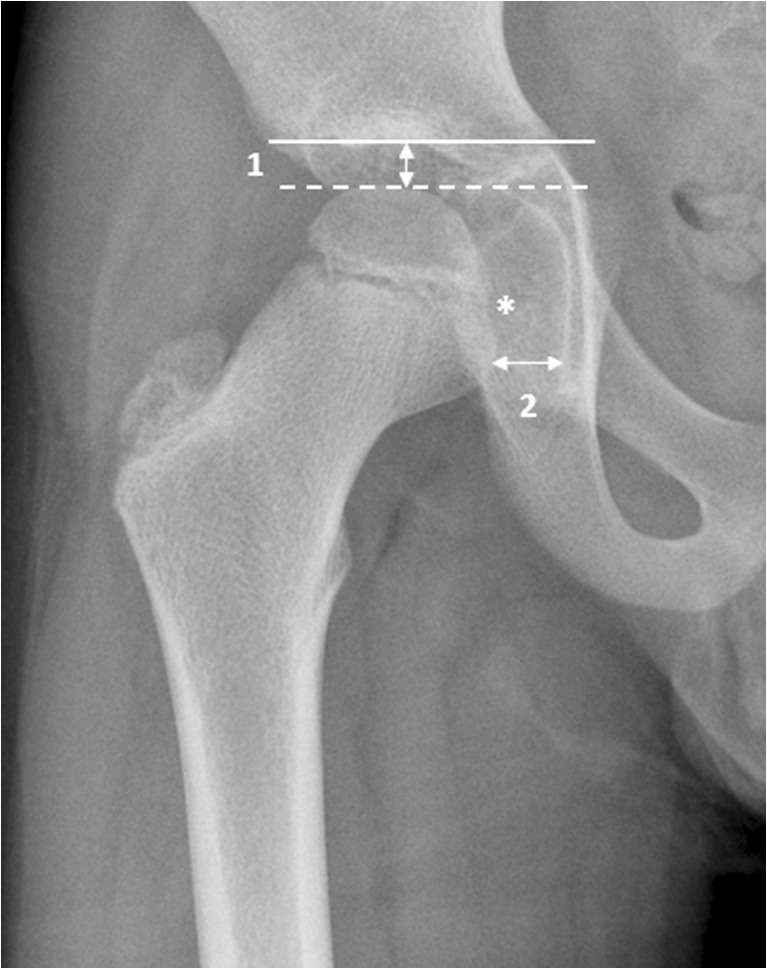
Fig. 3.
Demonstration of joint space measurements on an anteroposterior pelvic radiograph in a 9-year-old boy with juvenile idiopathic arthritis. We performed measurements in two locations: (1) superiorly, we drew a horizontal line along the roof of the acetabulum (solid line) and the most superior aspect of the femoral head (dashed line) and we calculated the distance between the two (arrow labelled 1); and (2) medially, we measured the joint space to the center of the femoral head (arrow labelled 2), just below the fovea (*)

