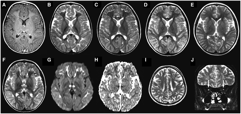
Figure 2.
Neuroimaging findings. In Subject 5, at age 3 years, axial T1-weighted post-contrast image (A) demonstrates hypointensity in the subcortical white matter, without abnormal enhancement. Axial T2-weighted images from the same patient at ages 3 (B), 6 (C), 7 (D) and 9 years (E) disclose progressive improvement of T2 hyperintense abnormalities in thalami, subcortical white matter and corpus callosum, initially appreciated in B. Axial T2-weighted image from the same patient at age 6 years (F) shows sparing of mammillothalamic tracts, seen as T2 hypointense dots in the centre of the signal change in the anterior portion of thalami. Considering the subsequent improvement, the restricted diffusion [hyperintensity in diffusion image (G) and hypointensity in apparent diffusion coefficients map (H)] probably reflects intramyelinic oedema. Axial and coronal T2-weighted images from Subject 3 at age 27 years (I and J), demonstrate residual T2 hyperintensity in subcortical white matter (I) and hypoplasia of both optic nerves (arrows in J).

