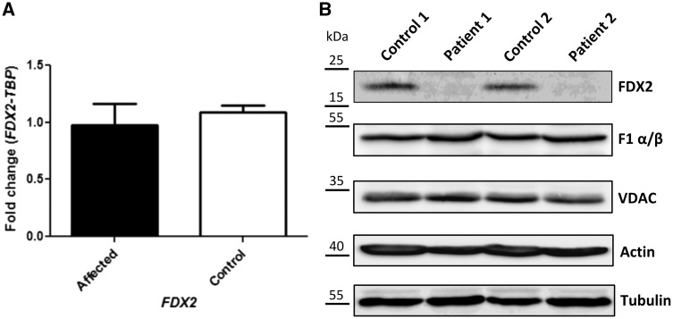
Figure 4.
Functional studies. (A) RT-qPCR showing relative FDX2 expression normalized to TBP in patients and controls. (B) Western blotting in patient and control muscle. Immunostaining was performed using specific antibodies against the mitochondrial protein FDX2, the F1 α/β subunits of complex V, and the voltage-dependent anion channel (VDAC, porin). Staining against actin and tubulin served as a loading control.

