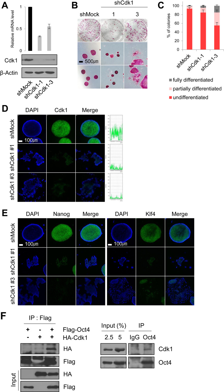
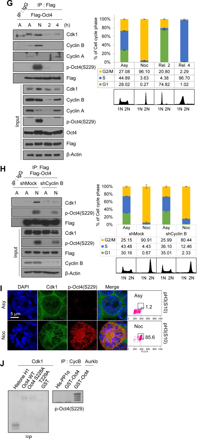
Figure 1.
Cdk1 is important for ESC pluripotency and preferentially interacts with Oct4 at the G2/M phase. (A) Protein levels and mRNA levels of Cdk1-knockdown mESCs (n = 3). (B and C) AP staining of Cdk1-knockdown mESCs. After staining to reveal AP activity, the colonies were scored and the percentages of undifferentiated, partially differentiated, and fully differentiated colonies were calculated. AP staining experiments were repeated three times (n = 3). Scale bar represents 500 μm. (D) Immunostaining of Cdk1-knockdown mESCs. Cdk1 was stained with anti-Cdk1 (green) and DNA was stained DAPI (blue). Scale bar represents 100 μm. (E) Fluorescence images of Cdk1-knockdown mESCs. Nanog and Klf4 were stained with anti-Nanog (green) and anti-Klf4 (green), respectively. DNA was stained with DAPI (blue). Scale bar represents 100 μm. (F) Flag-Oct4 and HA-Cdk1 were cotransfected into HEK293T cells. Cell lysates were immunoprecipitated with anti-Flag antibody and probed with anti-HA antibody (left). Endogenous Cdk1 was immunoprecipitated from E14 mESCs with anti-Oct4 antibody and immunoblotted with anti-Cdk1 antibody (right). (G) Changes in interaction of Cdk1 with Oct4 during cell cycle progression. Flag-Oct4-expressing ZHBTc4 mESCs were treated with nocodazole (100 ng/ml) for 8 h. At the indicated time after release into fresh media, cell cycle progression was determined by FACS analysis (right). Cell lysates were pulled down with anti-Flag beads. Bound proteins were eluted and then immunoblotted with the indicated antibodies. (A, asynchronous state; N, nocodazole treatment). (H) Changes in interaction of Oct4 with Cdk1 depending on Cyclin B knockdown. After Cyclin B knockdown, Flag-Oct4-expressing ZHBTc4 mESCs were treated with nocodazole for 8 h. Flag-Oct4 was immunoprecipitated by incubating lysates with anti-Flag beads. Following Flag elution, bound proteins were immunoblotted with the indicated antibodies. (I) Colocalization of Cdk1 and p-Oct4(S229) in mitosis-arrested mESCs was analyzed by immunostaining. mESCs were treated with nocodazole (100 ng/ml) for 8 h. Cdk1 was stained with anti-Cdk1 (green), p-Oct4(S229) was stained with anti-p-Oct4(S229) (red), and DNA was stained with DAPI (blue). Scale bar represents 5 μm. (J) Radioactive in vitro kinase assay using recombinant Cdk1 to phosphorylate GST-Oct4 wild type (WT) and S228A/S229A mutant. Autoradiogram showing incorporation of γ-32P ATP (left). For the IP kinase assay, cell lysates of nocodazole-treated E14 mESCs were immunoprecipitated with anti-CycB1 and Aurkb and subjected to an in vitro kinase assay using (His)6-PP1α and GST-Oct4 as a substrate and followed by western blotting (right).


