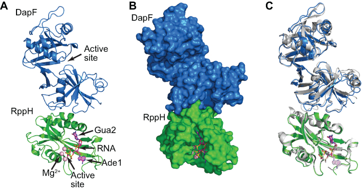
Figure 3.
Crystal structure of an RppHt-DapFm heterodimer bound to ppcpAGU RNA. (A) Overall view. The proteins are in cartoon representation, and the RNA ligand is in stick representation. RNA is colored violet, with nitrogen, oxygen and phosphorus atoms shown in their ‘atomic’ colors (blue, red and yellow, respectively). Mg2+ cations are depicted by light pink spheres. (B) Surface representation of the proteins, highlighting the shape complementarity of the RppH–DapF interface. (C) All-atom superposition of the RppHt-DapFm-RNA complex with isolated E. coli DapF and RppH-RNA structures (shown in grey color).

