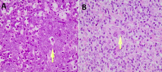
Figure 1.

Histopathological features of lymphomas after H and E staining: A) features seen in Burkitt's lymphoma, a NHL, were presence of histiocytes with phagocytosis of nuclear debris, accumulated cellular debris due to increased apoptosis, starry-sky pattern; B) features seen in lymphocyte predominance a HL, included inflammatory cell infiltrate composed of abundant eosinophils, plasma cells, and Reed-Sternberg cell; (Adapted from the University Teaching Hospital- Histopathology Laboratory)
