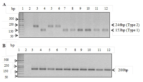
Figure 4.

A) EBV detection and subtyping targeting the EBNA 3C region; Lane 1, 50bp DNA marker; lane 2, negative control, lane 3 and 4, positive controls for type 2 and type 1, respectively; lanes 5-12 EBV-positive representative samples; B) actin internal control on selected samples. Lane 1, 50bp DNA marker; lane 2, Negative control; lane 3 and 4, positive controls for type 2 and type 1, respectively; lanes 5-12 representative samples
