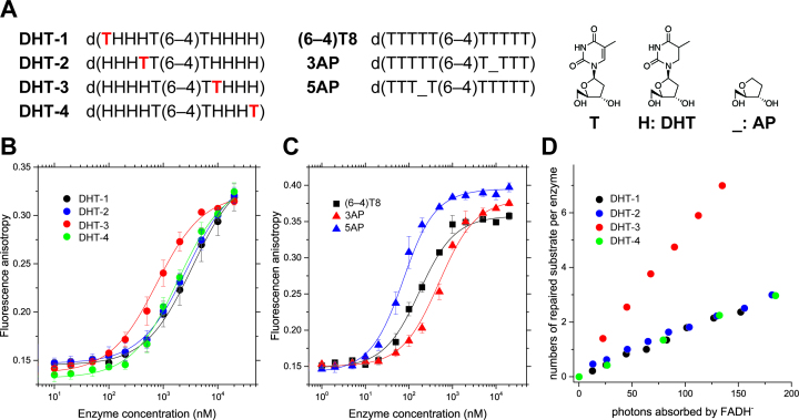
Figure 5.
Binding and photorepair properties of WT-Xl64 using chemically modified substrates. (A) Structures of the DHT and AP substrates. The 5′ terminus of each oligonucleotide for the anisotropy measurement was modified with the C3 amino linker on a DNA synthesizer, followed by labeling with TAMRA, while the DHT substrates used for the DNA repair assays were unlabeled, and had the intact 5′-hydroxy group. (B) Fluorescence anisotropy measurements of the fluorescently labeled DHT-1 (black), DHT-2 (blue), DHT-3 (red), and DHT-4 (green) upon titration with increasing amounts of WT-Xl64. The measurements were performed independently in triplicate, and the mean ± SD values are shown. (C) Fluorescence anisotropy measurements of fluorescently labeled (6–4)T8 (black square), 3AP (red triangle), and 5AP (blue triangle) upon titration with increasing amounts of WT-Xl64. (D) Photorepair of the DHT substrates by WT-Xl64.

