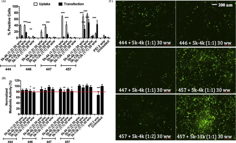
Figure 4.

Flow cytometry data showing uptake efficacy at 4 hrs and transfection efficacy at 2 days post treatment of H446 cells with 16 different formulations of PEG-PBAE polyplexes. The efficiency is in terms of percentage of live H446 cells positive for Cy3 (uptake) or EGFP (transfection). Data are mean ± SD (n=4)(*** p < 0.001, ** p < 0.01 compared to untreated). (B) Cytotoxicity of PEG-PBAE polyplexes, quantified by normalizing metabolic activity to untreated cells. Data are mean ± SD (n=3)(*** p < 0.001, ** p < 0.01, * P < 0.05 compared to untreated, red line marks 80% viability). (C) Representative fluorescence microscope images (10×) at 48 h post-treatment of H446 cells transfected with 6 different PEG-PBAE polyplex formulations. Scale bar is 200 μm for all panels.
