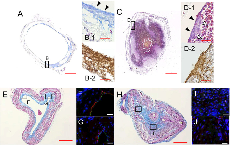
Fig. 4.

Representative histomorphological findings of BVGs. At day 1 after BVG implantation, a patent BVG (A, B-1; Carstairs’ Method, B-2; vWF antibody staining) showed a thin platelet-rich layer in the luminal side of BVG (B-1, arrowheads). An occluded BVG (C, D-1; Carstairs’ staining, D-2; vWF antibody staining) showed platelet-rich layers (D-1, arrowheads) and a large red thrombus in the luminal side of BVG (D-1, white arrowheads). The microscopic findings of the neovessel at 8 weeks are shown at which time the BVG scaffold had largely degraded. A patent neovessel showed a thick collagen layer (E; Masson’s Trichrome) and thin SMC layer (F, G; Immunofluorescent staining, Green; CD31, Red; a-SMA) in the luminal side. An occluded neovessel showed rich collagen and less SMCs (H-J). This finding was consistent with organized thrombi. Bar scale; 200 μm (A, C, E, H), 20 μm (B, D, F,G, I, J).
