
Figure 1. Inhibition of Osteoclastogenic Activity of Breast Cancer Cell CMs by mH2A1.2.
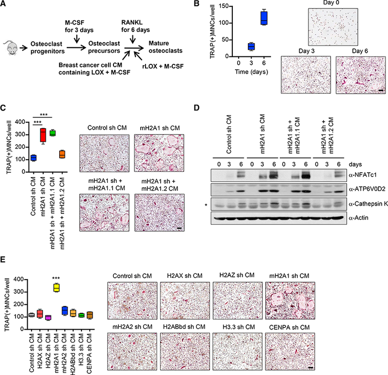
(A) Shown are the steps for analysis of osteoclastogenic properties of breast cancer cell-derived conditioned media (CMs) and recombinant LOX (rLOX).
(B) OCP cells were incubated with RANKL and MDA-MB-468 CMs for 0, 3, or6 days, fixed, stained for TRAP, and photographed under a light microscope (10×) (right). Representative images of osteoclasts are shown (scale bar, 100 μm). TRAP positive multinucleated cells (TRAP(+)MNCs) containing three or more nuclei and a full actin ring were counted as osteoclasts (left). Scale bar, 100 μm.
(C) CMs were prepared from mH2A1 -depleted MDA-MB-468 cells expressing exogenous shRNA-resistant mH2A1.1 or mH2A1.2 and analyzed for osteoclastogenic activity as in (B). Scale bar, 100 μm. Error bars are the means ± SD (n = 4) of three independent experiments; ***p < 0.001 (ANOVA analysis).
(D) Cell lysates were collected from OCP cells after treating with MDA-MB-468 CMs for the indicated time periods and analyzed for the expression of three osteoclast markers (NFATc1, ATP6V0D2, and cathepsin K) by western blot. β-Actin was used as a loading control. Non-specific band was marked by asterisk.
(E) OCP cells were cultured with CMs collected from MDA-MB-468 breast cancer cells depleted of the indicated histone variants. At the end of day 6, OCP- induced cells were stained for TRAP (right) to measure osteoclast differentiation (left). Error bars represent the means ± SD (n = 4); ***p < 0.001 versus Control sh CM (ANOVA analysis).