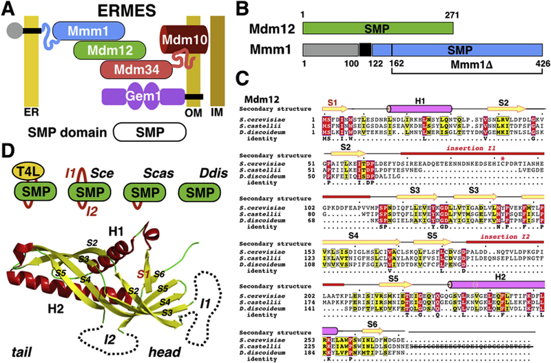
Fig. 1. ERMES and the SMP domain.

(A) Schematic of the yeast ERMES bridging the endoplasmic reticulum (ER) and mitochondria (Outer and Inner) membranes. (B) Domain organization of yeast Mdm12 and Mmm1. Mdm12 consists of an SMP domain while Mmm1 contains a luminal domain (grey), one transmembrane anchor and a single cytoplasmic SMP domain (blue). (C) Protein sequence alignments of Mdm12 in Saccharomyces cerevisiae, Saccharomyces castellii and Dictyostelium discoideum. Non-conserved insertions (I1 and I2) are highlighted. Secondary structure elements are labeled. (D) Two variable insertions in the SMP fold of Mdm12: I1 (absent in Scas and Ddis) and I2 (absent in Ddis). The T4L insert replaces the longest insertion I1. (For interpretation of the references to colour in this figure legend, the reader is referred to the web version of this article.)
