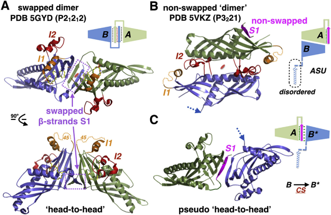
Fig. 3. A new crystal structure of Sce-Mdm12.

(A) Swapped ‘head-to-head’ dimer of Sce-Mdm12 observed in the 3.1 Å resolution orthorhombic structure from Jeong et al. [22]. (B) Non-swapped dimer of Sce-Mdm12 observed in our 4.1 A resolution rhombohedral structure. Two monomers (A and B) are observed in the asymmetric unit. In monomer A, the N-terminal β-strand S1 (magenta) is resolved in the electron density but does not swap. The N-terminal β-strand S1 of monomer B has flipped into a solvent-exposed conformation and cannot be resolved in the electron density maps. Insertions I1 and I2 are colored in gold and red, respectively. (C) A crystallographic symmetry-related copy of monomer B (note B*), forms a pseudo ‘head-to-head’ dimer where only one β-strand S1 (from monomer A) sits at the interface between the two SMP/TULIP domains. CS indicates that B and B* are related by a P3221crystallographic symmetry operator. (For interpretation of the references to colour in this figure legend, the reader is referred to the web version of this article.)
