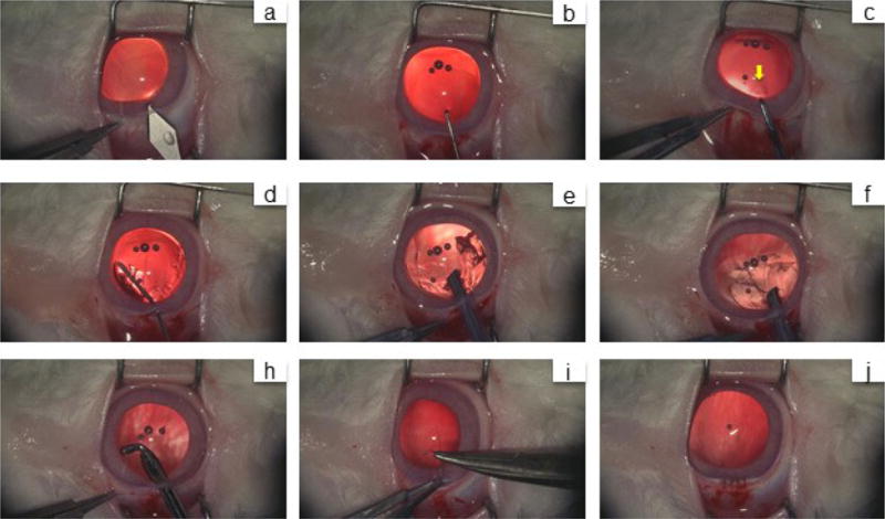
Extended Data Figure 6. Lens regeneration surgery in rabbits.

a, A 3.2-mm keratome was used to make a limbus tunnel incision at the 11-12 o’clock position into the anterior chamber. b, The capsular opening was created by a capsulorhexis needle. c, A 1-2 mm diameter anterior capsulotomy was performed using the anterior continuous curvilinear capsulorhexis (ACCC) technique near the capsular opening area (yellow arrow). d, A blunt needle was used to inject balanced salt solution for hydrodissection of the cortex from the anterior capsule. e, The cortex was removed using a phacoemulsification device. f, The remaining cortex was removed using irrigation and aspiration. h, An elbow I/A handle was used to clear the equatorial cortex. i, j, The limbus wound was sutured with an interrupted 10-0 nylon suture. The wound was found to be watertight.
