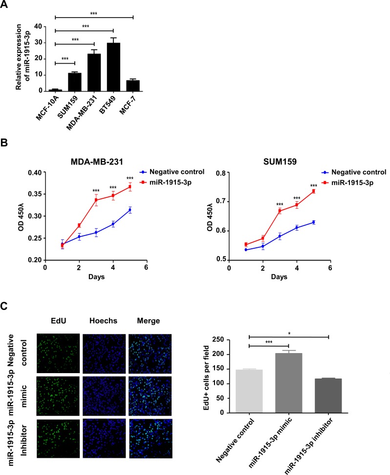
Fig 4. miR-1915-3p promoted breast cancer cell proliferation in vitro.
(A) miR-1915-3p expression in breast cancer cells were measured by Taqman probe RT-qPCR assay. (B) CCK8 assay was performed on both MDA-MB-231 and SUM-159 cells which were transfected with miR-1915-3p mimics (red line) or negative control (blue line). OD450 value was measured every 24 hours during 5 days culture. (C) The EdU staining (Green) of MDA-MB-231 cells transfected with miR-1915-3p mimic or inhibitor was performed after seeding cells 72 hours, meanwhile cell nucleus were stained with Hochest (Blue), statistic result was shown on the right. Data was shown as mean±s.e.m. from three independent experiments (***P<0.001,**P<0.01, *P<0.05).

