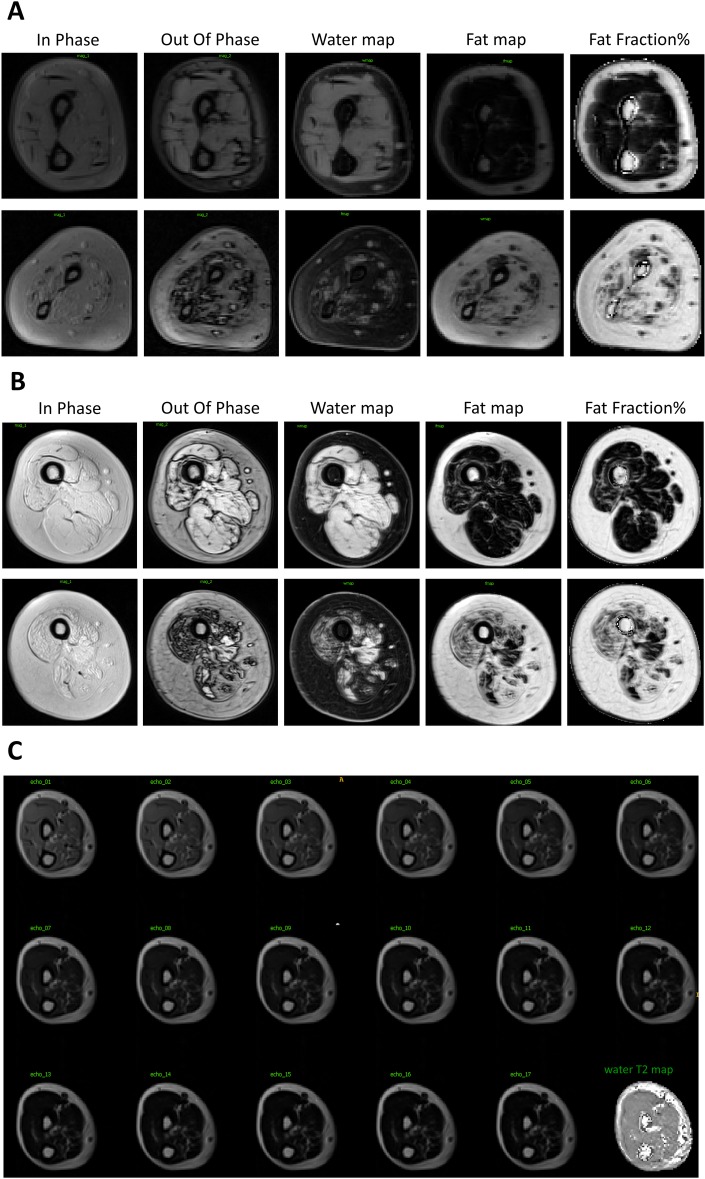
Fig 3. Example of MRI images of patients with SMA type 2 and 3.
A. Example of Dixon images and water T2 map in mildly (upper panel; non-ambulant SMA type 3) and more severely (lower panel; non-sitter SMA type 2) infiltrated patient forearms; B. Example of Dixon images and water T2 map in mildly (upper panel) and more severely (lower panel) infiltrated ambulant patient thighs; C. Example of series of spin echo images at increasing TEs (10 ms steps) in the forearm of a patient and the water T2 map reconstructed from this series using the tri-exp fitting method.

