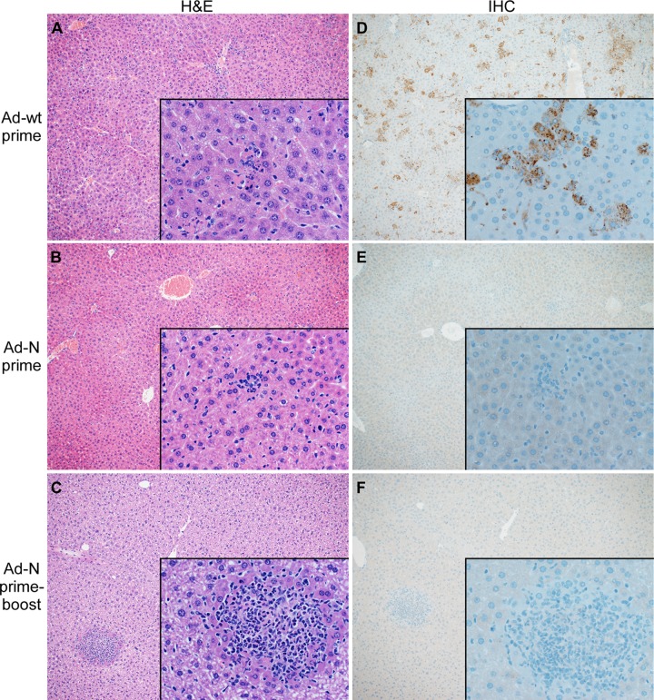
Fig 3. Liver histopathology and CCHFV antigen distribution in single-dose and prime-boost vaccinated and challenged mice.
Groups of IFNAR-/- mice were either single, (1.25×10⁷ IFU; intramuscular) or prime-boost (1.25×10⁷ IFU; intramuscular / 108 IFU; intranasal) vaccinated with Ad-N or Ad-wt and challenged with 1000 LD50 of CCHFV 28 days following final vaccination. Mice (n = 9 per group) were anesthetized, bleed and euthanized to harvest organ samples on day 3 post CCHFV challenge. Thin-sections of liver material were stained with hematoxylin and eosin (H&E) or with N1028 rabbit polyclonal serum (anti-CCHFV N serum) (IHC). (A) Liver H&E of control-vaccinated mice (Ad-wt), (B) Liver H&E of prime-vaccinated mice (Ad-N); (C) Liver H&E of prime-boost-vaccinated mice (Ad-N); (D) Liver IHC of control-vaccinated mice (Ad-wt); (E) Liver IHC of prime-vaccinated mice (Ad-N); (F) Liver IHC of prime-boost-vaccinated mice (Ad-N). Images are at a magnification of 10x with 500x insets.

