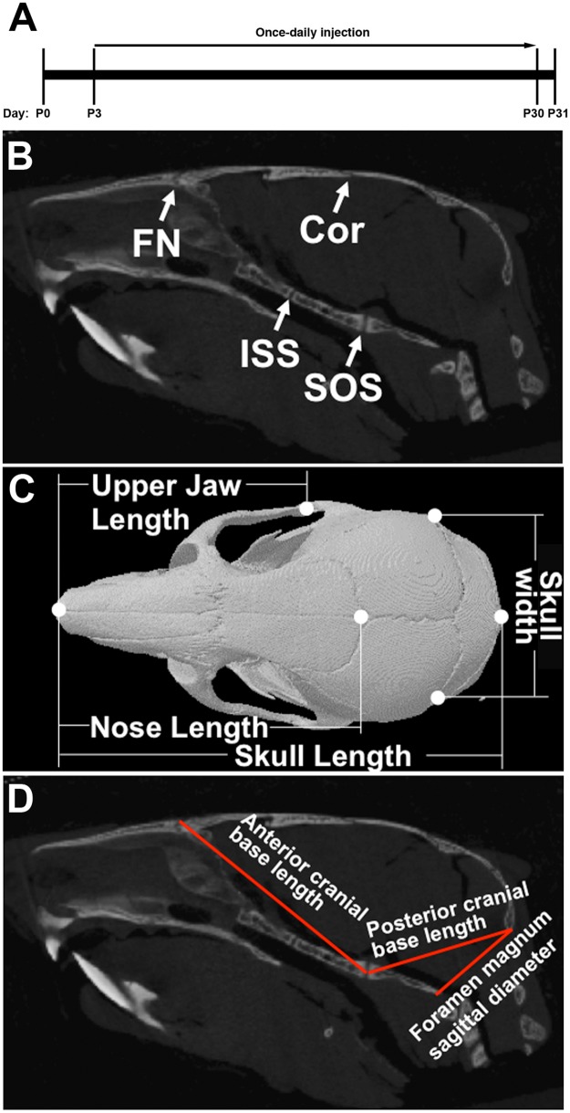
Fig 1. Locations of sutures and linear dimensions analyzed in the skull.
(A) BMN 111 treatment timeline. (B) 2D sagittal slice of μCT data showing the coronal (Cor) and frontonasal (FN) sutures and the intersphenoidal (ISS) and spheno-occipital (SOS) synchondroses. (C) 3D reconstruction of μCT data showing a dorsal view of the skull with skull, nose, and upper jaw lengths and skull width indicated. (D) 2D sagittal slice of μCT data showing the anterior and posterior cranial base lengths and the foramen magnum sagittal diameter.

