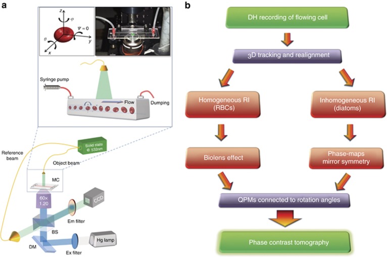
Figure 1.
Working principles of the R-TPM approach. (a) Sketch of the experimental R-TPM set-up. Cells are injected into a microfluidic channel and tumble while flowing along the y-axis (inset of a). At the same time, a holographic image sequence is acquired. In the top-left corner of the inset, the reference system for cell tumbling is reported; in the top-right corner, a photo of the real set-up is shown. Rotation occurs around the x and z axes. BS, beam splitter; DM, dichroic mirror; MC, microchannel. (b) Flow chart representing the main steps of the two proposed algorithms for angle recovery and tomographic reconstruction.

