. 2016 Aug 3;5(6):1604–1618. doi: 10.1039/c6tx00225k

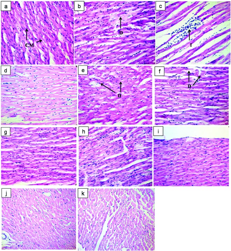
Fig. 6. Impact of resveratrol and pterostilbene on the histopathological features of heart tissue in the AEST administered rats. Representative photomicrographs of haematoxylin and eosin (H and E) stained sections of cardiac tissue: (a) vehicle control showing healthy cardiac myocytes, suggesting normal cardiac physiology including structure and function; (b) VCD (80 mg kg–1, i.p.) administered rats showed moderate degenerative changes in cardiac myocytes; (c) AEST (100 mg kg–1 p.o.) administration causes inflammatory changes with decreasing density of cardiac myocytes; (e) RSV 5 (5 mg kg–1, p.o.), (h) PT 5 (5 mg kg–1 p.o.) treated rats showed mild degeneration of cardiac muscle cells; importantly, (d) ET (0.05 mg kg–1, s.c.), (f) RSV 20 (20 mg kg–1, p.o.), (g) RSV 40 (40 mg kg–1, p.o.), (i) PT 20 (20 mg kg–1, p.o.), (j) PT 40 (40 mg kg–1, p.o.) and (k) CT (RSV 20 + PT 20 mg kg–1, p.o.) treatment showed no signs of cardiac damage [H and E ×400]. Abbreviations: CM: cardiac myocytes; D: degenerative change in cardiac myocytes; I: infiltration of leucocytes.

Fig. 6