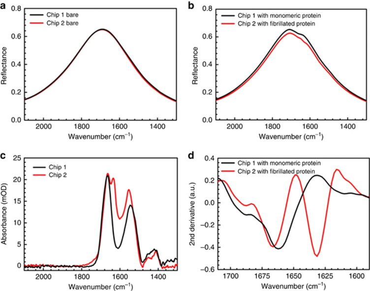
Figure 2.
Spectroscopic detection of secondary structure components of monomeric and fibrillated α-Syn protein on plasmonic biosensors. (a) Bare reflectance response of clean antenna arrays with L: 2200 nm and P: 2.85 μm on two different substrates: an array on Chip 1 in black used for the monomeric α-Syn experiment and an array on Chip 2 in red used for the fibrillated α-Syn experiment. (b) Responses of the same antenna arrays shown in (a) after physisorption of monomeric α-Syn on Chip 1 (black) and fibrillated α-Syn on Chip 2 (red), followed by rinsing. (c) Extracted absorbance signals for monomeric (black) and fibrillated (red) α-Syn protein with baseline correction. (d) Second derivatives of the acquired logarithmic reflectance ratios (A) from monomeric (black) and fibrillated (red) α-Syn, showing the distinguished β-sheet signal in the aggregated samples.

