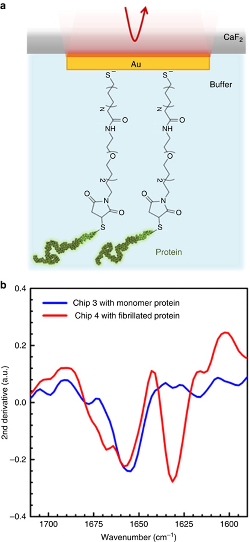
Figure 3.
Spectroscopic detection of secondary structure components of monomeric and fibrillated α-Syn protein immobilized on plasmonic biosensors with SAM in buffer solution. (a) Schematic illustration of the in-solution measurement configuration from the backside of the CaF2 substrate (not to scale), and the protein immobilization with MHDA/amino-(PEG)2-maleimide linker. (b) Second derivatives of the acquired amide I signals measured in Tris buffer from monomeric (blue) and fibrillated (red) α-Syn, immobilized on Chips 3 and 4, respectively (similar antenna designs with L: 1850 nm and P: 2.6 μm), showing the distinguished β-sheet signal in the aggregated samples compared to random structured monomers.

