. 2017 Aug 24;6(6):878–888. doi: 10.1039/c7tx00126f

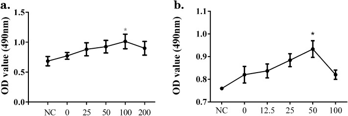
Fig. 1. Supernatants from silica-treated macrophages induced the proliferation of NIH-3T3 and MRC-5 cells. Raw264.7 cells were treated with different concentrations of silica for 24 h and the supernatants were used to culture fibroblasts. NIH-3T3 cells were cultured in DMEM (NC) or the supernatants collected from Raw264.7 cells exposed to 0, 25, 50, 100 or 200 μg mL–1 silica solution for 24 h. MRC-5 cells were cultured in DMEM (NC) or the supernatants collected from Raw264.7 cells exposed to 0, 12.5, 25, 50 or 100 μg mL–1 silica solution for 24 h. The MTT assay was performed to measure cell proliferation. In the case of NIH-3T3 (a), the peak value and significant proliferation compared with the NC group were shown in the silica 100 μg mL–1 group, while for MRC-5 (b) these appeared in the silica 50 μg mL–1 group. The results show data from five repeated tests. The data are presented as means ± SD. The significance of differences was determined by ANOVA followed by the Dunnett's test; *p < 0.05 versus the NC group.

Fig. 1