. 2017 Aug 24;6(6):878–888. doi: 10.1039/c7tx00126f

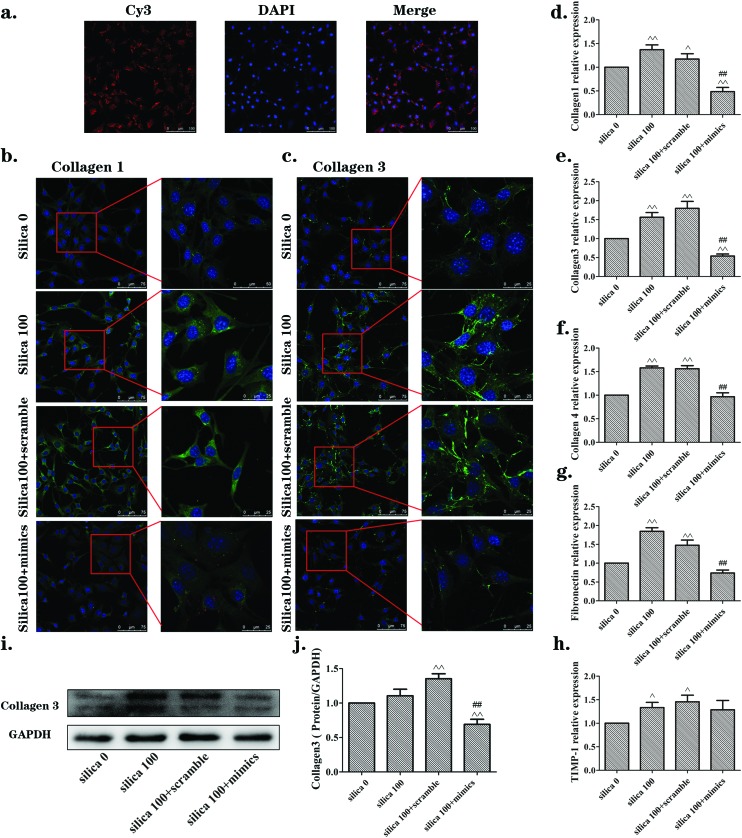
Fig. 5. The miR-29b inhibited the activation of NIH-3T3 cells induced by supernatants from silica-treated macrophages. (a) Transfection efficiency for NIH-3T3 cells checked by examination of Cy3 through laser scanning confocal fluorescence microscopy after 24 h incubation. (b, c) Immunofluorescence staining of collagen1 and collagen3 (green), respectively. Levels were obviously increased in silica 100 μg mL–1 and silica 100 + scramble groups, but were significantly reversed in the silica 100 + mimics group. (d, e, f, g, h) qRT-PCR results of mRNA expression levels of collagen 1, collagen3, collagen4 and fibronectin, respectively. mRNA levels were increased following induction by silica, while they were reversed in the silica 100 + mimics group. (i, j) Western blotting analyses of the protein expression levels of collagen3. The collagen3 protein levels were consistent with the qRT-PCR results. The data are presented as the means ± SD (n = 3). The significance of differences was determined by ANOVA followed by the Dunnett's test; ^p < 0.05 versus the silica 0 group, ^^p < 0.01 versus the silica 0 group, #p < 0.05 versus the silica 100 group, ##p < 0.01versus the silica 100 group.

Fig. 5