. 2016 Mar 16;5(3):931–937. doi: 10.1039/c5tx00471c

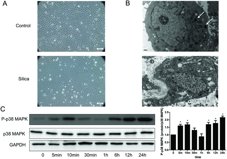
Fig. 4. Silica changed the cell morphology, ultrastructure and activated the p38 MAPK signaling pathway in RLE-6TN cells. (A) Inverted microscope analysis of the RLE-6TN cells cultured under different conditions. A typical epithelial cuboidal shape of the RLE-6TN cells in the control group was shown, with the characteristic cobblestone morphology, while the cells became more elongated, less adhered and lost their apical-to-basal polarity after treatment with the 50 μg per ml silica supernatant. (B) Ultrastructure analysis of RLE-6TN cells. Cells in the control group showed multiple mature lamellar bodies and microphilopodia, while in the silica group there was substantial reduction of mature lamellar bodies. MLB: mature lamellar bodies. (C) P-p38 MAPK and p38 MAPK were measured by western blot of the RLE-6TN cells cultured with or without silica for 48 h. *P < 0.05 vs. control.

Fig. 4