. 2016 Jan 11;5(2):583–593. doi: 10.1039/c5tx00305a

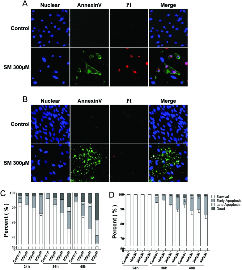
Fig. 2. Effects of SM on mode of cell death (necrosis, apoptosis) in HEK-f and HDF-a cells. A and B: representative images of the nucleus, cell membrane eversion (annexinV), and nuclear necrosis (PI) in HEK-f cells and HDF-a cells, respectively. C and D: effects of different concentrations of SM on cell necrosis and apoptosis at different treatment times in HEK-f cells and HDF-a cells, respectively. The nucleus was stained with Hoechst 33342. Cell membrane eversion was detected using phosphatidylserine (PS)-labeled annexinV. Nuclear necrosis was stained with propidiumiodide (PI). The percentage of cells in four groups (survival, early apoptosis, late apoptosis, necrosis) was derived from the two parameters cell divide analysis method (Linear Discriminant 2D Filter in the Multi Target Analysis Module, GE Health Care, USA) according to the intensity of annexinV and PI staining on images. All the results were normalized and expressed as percentage changes of the control. The data were expressed as mean ± standard error. All experiments were performed in triplicate, with no less than 200 cells per well. A measured difference of 3 SDs (standard deviation) was considered a significant change.

Fig. 2