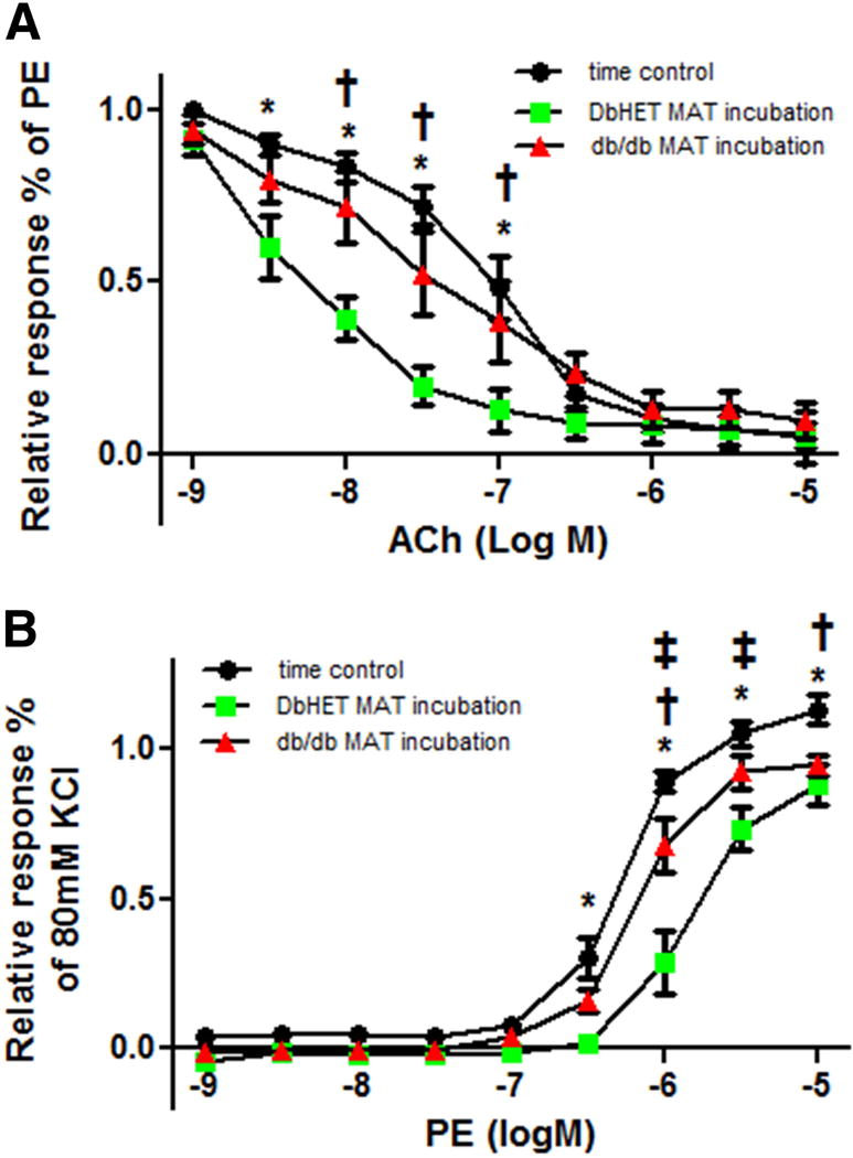
Figure 8. Acute incubation of DbHET arteries with db/db PVAT impairs vasoreactivity.

Panel A: Incubation of DbHET MA with PVAT from the same animal increased sensitivity to ACh, relative to time controls (as shown in Figures 6 and 7). In contrast, the increase in sensitivity was not observed when DbHET MA was acutely (1 hour) incubated with PVAT from db/db mice. Panel B: Incubation of DbHET MA with PVAT from the same animal decreased sensitivity to PE, relative to time controls. In contrast, the decrease in sensitivity was not observed when DbHET MA was acutely (1 hour) incubated with PVAT from db/db mice. logEC50 values are provided in Supplementary Table 1. Data presented as mean± SEM. N=6 per group. Concentration-response curves were analyzed by Two-way ANOVA. *: P < 0.05. time control vs DbHET MA+ MAT from DbHET mice. †: P < 0.05. time control v.s. DbHET MA+ MAT from db/db mice. ‡: P < 0.05. DbHET MA+ MAT from DbHET mice v.s. DbHET MA+ MAT from db/db mice.
