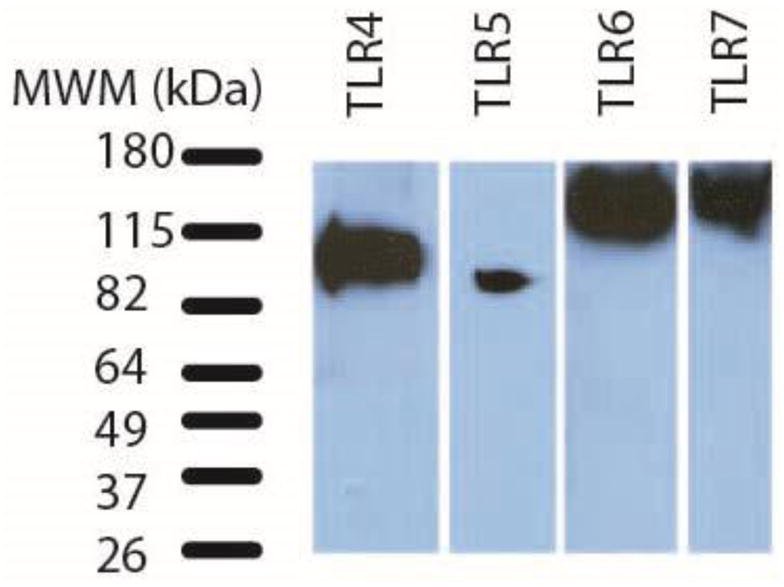
Figure 1. TLRs 4-7 are detected in macaque neural retina tissue lysates by immunoblotting.

Immunblots of macaque retina tissue lysates demonstrate expression of TLR 4-7 proteins. Table 2 lists amount of retina lysate loaded and immunoblotting conditions for each antibody. This figure is a composite of 4 separate immunoblots. The bands in the MWM lane represent the migration of BenchMark Pre-Stained Protein Standard on a 4–15% Mini-PROTEAN TGX precast gel.
