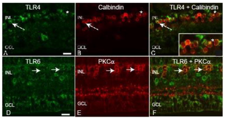
Figure 8. TLRs 4 and 6 are expressed by bipolar cells in the macaque retina.
A) The arrow and asterisk indicate TLR4 staining of round cells in the INL of macaque retina. B) The arrows and asterisk indicate calbindin positive OFF bipolar cells. C) The merged image indicates co-localization of TLR4 and calbindin positive cells. D) The arrows indicates TLR6 staining of round INL cells. E) The arrows indicates PKC-α positive rod bipolar cells. F) Merged image indicates co-localization of TLR6 and PKC-α stained cells. Inner nuclear layer (INL), ganglion cell layer (GCL). Scale bar= 20 μm.

