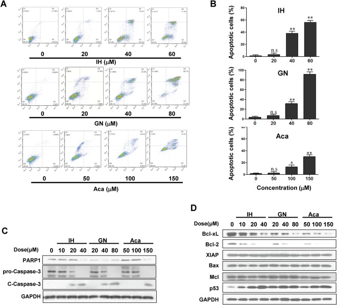
Figure 4.
IH, GN and Aca induce apoptosis in MDA-MB-231. MDA-MB-231 cells were treated with IH, GN, Aca at indicated concentrations for 48 h. (A,B) The percentage of apoptotic cells were determined by flow cytometry using Annexin V/PI staining for three independent experiments. (mean ± SD, n.sP > 0.05, *P < 0.05 or **P < 0.01 vs. the control). (C,D) Cell lysates were prepared and analyzed by western blotting using antibodies against PARP1, pro-caspase-3, cleaved-caspase 3 (C-Caspase 3), Bcl-2, Bcl-xL, p53, Bax, XIAP and Mcl-1.

