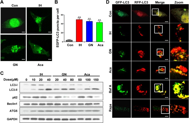
Figure 5.
IH, GN and Aca induce autophagy in MDA-MB-231. (A,B) MDA-MB-231 cells transfected with EGFP-LC3 were treated with 40 μM IH, 20 μM GN and 100 μM Aca for 24 h, the EGFP-LC3 puncta were observed under confocal microscopy; scale bars: 10 μm. Quantification of average EGFP puncta per cell for three independent experiments. Data was presented as mean ± SD (**P < 0.01 vs. the control). (C) Cells were exposed to indicated concentrations of IH, GN and Aca for 24 h, the expression of autophagy-related proteins (LC3B-II, p62, Beclin1, ATG5) were detected by western blot analysis, GAPDH was used as a loading control. (D) Cells were transfected with a tandem reporter construct (tfLC3), and were exposed to 30 μM IH, 20 μM GN, 100 μM Aca, 20 nM Baf and 0.25 μM Rapa as indicated. The colocalization of EGFP and mRFP-LC3 puncta was examined by confocal microscopy. Scale bars: 10 μm.

