Figure 2.
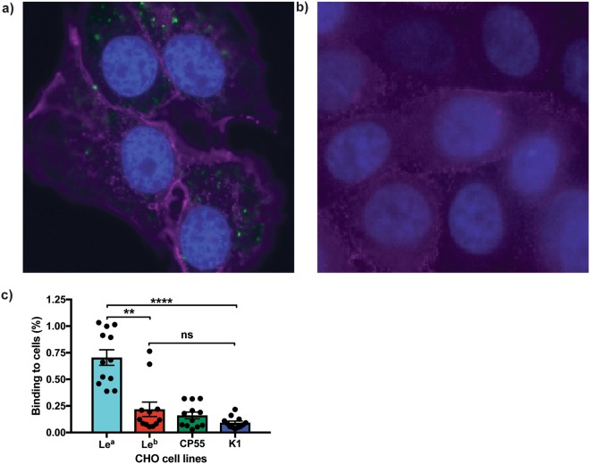
ETEC CFA/I fimbriae bind to the CHO-Lea cells. Immunofluorescence staining of (a) CHO-Lea and (b) CHO-Leb cells infected with TOP10-CFA/I. Infected cell lines were stained with anti-CFA/I antibody (Alexa Fluor 488, green), Lea antibody (Alexa Fluor 647, purple), Leb antibody (Texas Red, purple) and host nuclei stain (DAPI, Blue). All images are taken at x40 magnification. (c) TOP10-CFA/I bacteria adhere to the CHO-Lea cell line more than to CHO-Leb, CHO-CP55 and CHO-K1 cells. Graphs represent the percentage of TOP10-CFA/I bacteria binding to each of the cell lines, as measured by quantifiable immunofluorescence analysis. Statistical analysis was performed using ANOVA with Dunn’s multiple comparisons test. **Indicates P = 0.0065, ****Indicates P = <0.0001. Data presented as Mean and SEM of at least three independent experiments.

