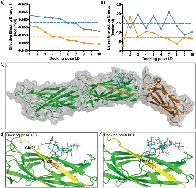
Figure 5.
Molecular docking predicts CfaB can bind to Lea-5 and Leb-6 glycans. (a) Effective Binding Energy I (kcal/mol) of the ten highest ranked Lea-5 and Leb-6 poses. (b) Linear Interaction Energy (kcal/mol) of the ten highest ranked Lea-5 and Leb-6 poses. Blue lines are Lea-5 poses and orange lines are Leb-6 poses. Dotted lines are respective mean values for the ten highest ranked Lea-5 (blue) and Leb-6 (orange) poses analysed. (c) Overall surface view and cartoon representation of Lea-5 binding to the N-terminal Ig-like groove of the major CFA/I subunit CfaB. (d) Cartoon representation of docking pose a03, showing hydrogen bond interaction of residues Glu25 and Thr29 of the CfaB Ig-like groove with Lea-5. (e) Cartoon representation of docked complex b01, showing hydrogen bond interaction of Thr29 of the CfaB Ig-like groove with Leb-6. For (c) to e) the Lea-5 and Leb-6 ligands are stick representations (cyan) with atoms interacting with CfaB coloured in yellow and hydrogen bonds displayed as black dotted lines. For the CFA/I fimbriae, the minor subunit CfaE is the green ribbon, the major subunit CfaB is the dark yellow ribbon and the CfaB N terminal Ig-like groove is the bright yellow ribbon.

