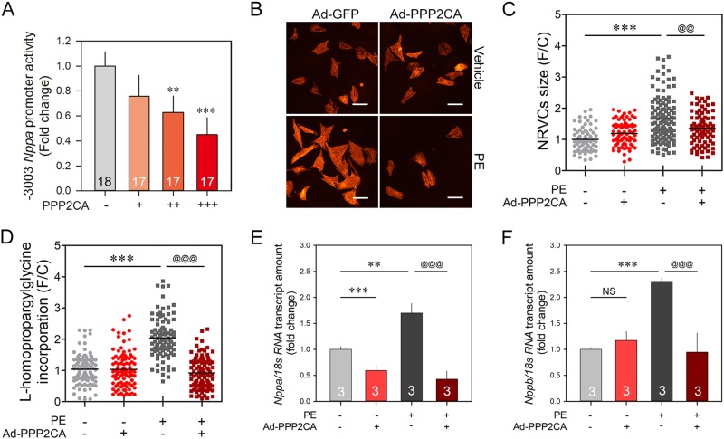
Fig. 4. PPP2CA negatively regulates hypertrophic responses.
a –3003 Nppa reporter activity in H9c2 cells was decreased in a PPP2CA dose-dependent manner. b, c Cell size measurement in NRVCs. Cardiomyocyte-specific sarcomeric alpha-actinin was visualized by immunocytochemistry analysis. Treatment with 20 μmol/L PE induced an increase in the cell surface area, a hallmark of cardiomyocyte hypertrophy. Ad-PPP2CA abolished the PE-induced cell size enlargement. The dots depict individual cell size. d De novo protein synthesis was evaluated by quantification of the incorporation of l-homopropargylglycine (HPG) in NRVCs. NRVCs were infected with Ad-PPP2CA for 48 h and exposed to 20 μmol/L PE. Cells were cultured in HPG-containing methionine-free medium for 1 h, and then the incorporated HPG was ligated to Alexa Fluor 594 azide for visualization. The signal density of the cells was determined by fluorescence microscopy and then quantified using software. Ad-PPP2CA significantly attenuated de novo protein synthesis driven by PE. e, f PE-induced early hypertrophic marker gene expression was regulated by Ad-PPP2CA. Both Nppa (e) and Nppb (f) were successfully increased by PE stimuli, which was blocked by infection of Ad-PPP2CA. Numbers in the bars (a, e, f) indicate the experimental set. White scale bars indicate 15 μm. @@ indicates p < 0.01. *** and @@@ depict p < 0.001

