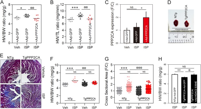
Fig. 7. PPP2CA negatively regulates cardiac hypertrophy through HDAC2 dephosphorylation in the heart.
a, b The anti-hypertrophic effect of PPP2CA was tested in CD1 mice. An in vivo hypertrophy model was tested using ISP infusion. Ad-PPP2CA was delivered via the tail vein 7 days after implantation of an ISP micro-osmotic pump, and the mice were sacrificed 14 days after the operation. Simultaneous expression of PPP2CA blunted ISP-induced cardiac hypertrophy, as determined either by heart weight per body weight ratio (HW/BW) (a) or by heart weight per tibia length ratio (HW/TL) (b). Dots depict individual mouse data. The mouse numbers used in a and b are as follows: 7 (Sham + Ad-GFP), 10 (ISP + Ad-GFP), and 8 (ISP + Ad-PPP2CA). c Quantitative real-time PCR revealed that Ad-PPP2CA successfully infected the myocardium. d Representative gross images. e–g Cardiac hypertrophy was significantly attenuated in TgPPP2CA mouse heart. Masson’s trichrome staining. Interstitial fibrosis induced by ISP infusion was dramatically reduced in TgPPP2CA mice (e). Transgenic expression of PPP2CA allowed resistance against the hypertrophy stimulus induced by infusion of ISP (f). Cross-sectional area in the left ventricle free wall showed a pattern similar to the HW/BW or HW/TL results (g). The anti-hypertrophic effect of PPP2CA against ISP infusion was not observed when Ad-HDAC2 S394E was expressed simultaneously in the heart of TgPPP2CA mice (H). * and @ depict p < 0.05. ** and @@ indicate p < 0.01. *** and @@@ mean p < 0.001. NS not significant

