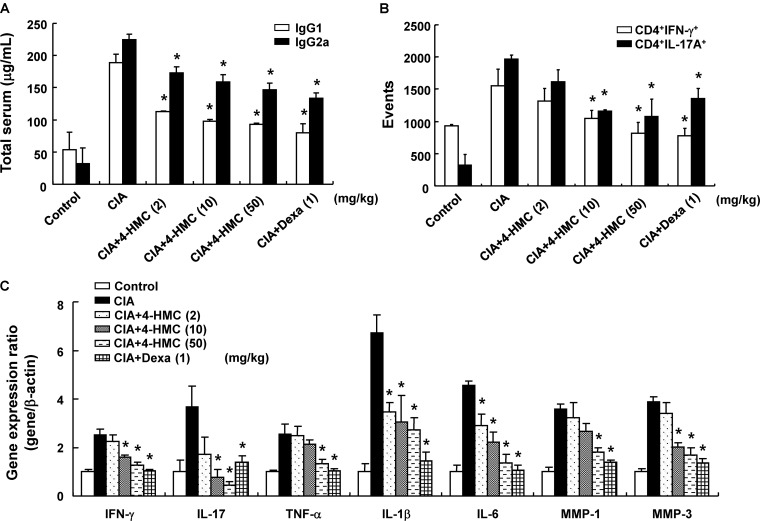
FIGURE 3.
Serum IgG levels, phenotypes of T cells in the lymph nodes, and cytokine expression in the joints of collagen-induced arthritis (CIA) mice. (A) Total serum IgG levels were measured by ELISA. (B) Inguinal lymph nodes were collected from each mouse, and the numbers of CD4+IFN-γ+ and CD4+IL-17A+ isolated single cells were detected using FACSCalibur flow cytometry. (C) The joints were excised, total RNA was isolated, and qPCR gene expression analysis was performed. The data are presented as the mean ± SD of five determinations. ∗p < 0.05, significantly different from untreated CIA mice. 4-HMC, 4-(hydroxymethyl)catechol; Dexa, dexamethasone.

