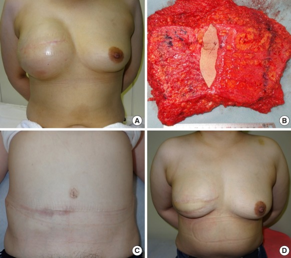
Fig. 4. The clinical course of case 4.

(A) Preoperative chest view after full expansion. (B) Transverse rectus abdominis myocutaneous (TRAM) flap view after harvest. It was partially de-epithelialized and zone IV was excised. (C) The view of the donor site at postoperative month 6. (D) Photograph showing symmetrical breasts at postoperative month 6.
