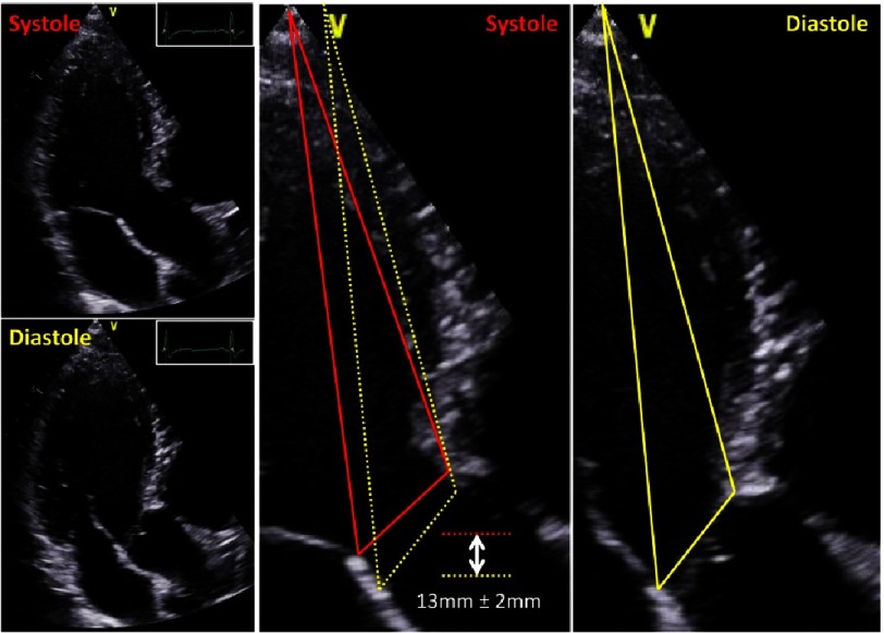
Figure 10. Caudal-cranial aortic annulus excursion between systole and diastole.
Due to LV filling during diastole the distance between apex and AV-junction is more cranial in comparison to end-systole. Changes of aortic annulus position are illustrated by yellow lines during diastole and red lines during systole.

