Figure 3.
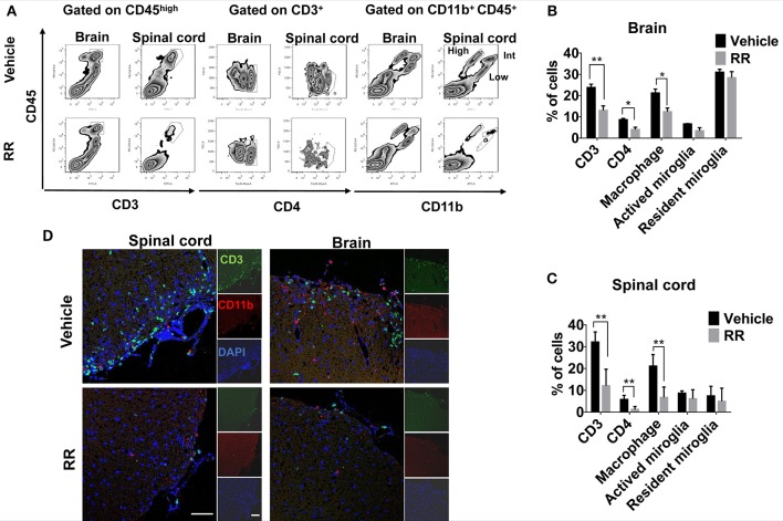
RR decreased the populations of CD3 + and CD11b + cells in active EAE. MNCs were isolated from brain or spinal cord in RR or vehicle-treated EAE mice at 30 dpi (treatment protocol). (A) Cells were analyzed for expression of CD3 or CD4 in the lymphocyte gate and that of CD11b in total MNC gate by flow cytometry. CD11b+ CD45high cell was defined as macrophage, CD11b+ CD45int as active microglia and CD11b+ CD45low as resident microglia. Percentages of positive cells in brain (B) or spinal cord (C) are represented (n = 4). (D) Immunofluorescent co-staining of CD3+(green) and CD11b+(red) cells with nucleus (blue) in spinal cord (left) and brain (near choroid plexus within lateral ventricle, right) at 30 dpi (scale bar, 50 μm). *P < 0.05, *P < 0.01.

