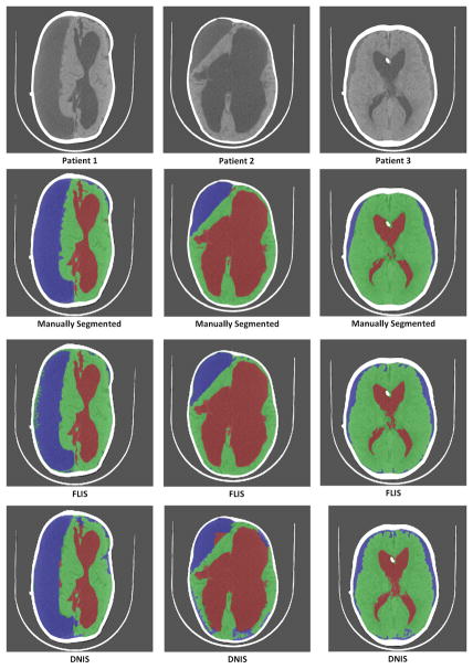
Fig. 16.
Comparison of results between DNIS and FLIS for training-test configuration of 17–15. First row represents the original images of 3 patients. Second row represents their corresponding manually segmented image. Third row represents segmented images using FLIS. Fourth row represent segmented images using DNIS. Green-Brain, Red-CSF, Blue-Subdurals.

