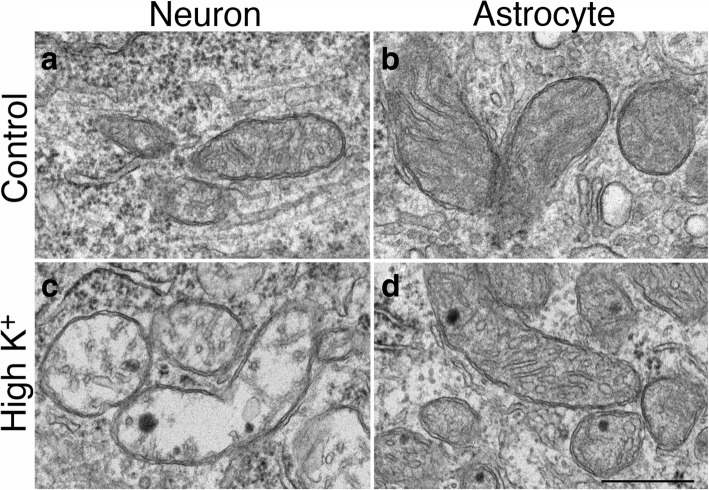
Fig. 10.
Mitochondria in neuronal somas became swollen upon depolarization (c vs. a; c was treated with 90 mM K+ for 3 min), while mitochondria in astrocytes displayed similar features under control conditions (b) or upon depolarization (d). Images of neurons and astrocytes were collected from the same samples of hippocampal slice cultures. Scale bar = 0.5 μm

