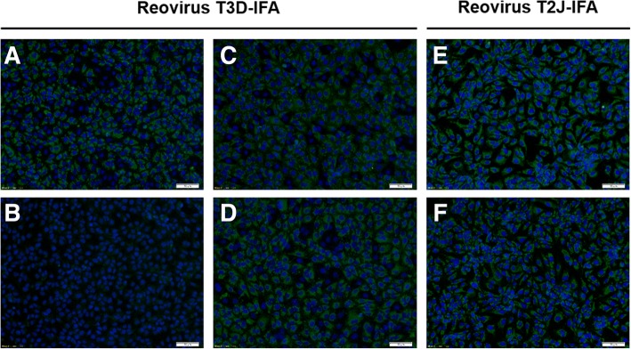
Fig. 2.
IFA confirms seroconversion of reovirus T3D-infected mice. IFA based on reovirus T3D- (a-d) and reovirus T2J-infected (e + f) MA104-cells probed with anti-reovirus type-3 σ-1 monoclonal antibody (mAb, a; positive control), serum of an uninfected specific pathogen-free (SPF) mouse (b; negative control) and sera of reovirus T3D-infected BALB/c (c + e) and C57BL/6 (d + f) mice. Fluorescence staining relied on an Alexa Fluor 488-conjugated goat anti-mouse IgG antibody (green). Cell nuclei were stained with DAPI (blue). Scale bar: 50 μm

