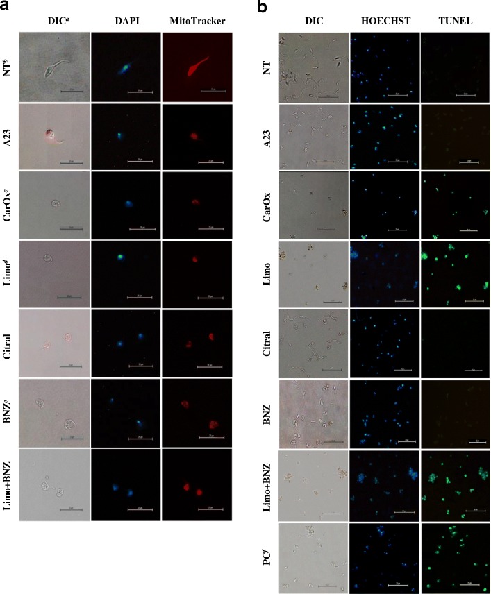
Fig. 4.
Cell morphology changes of Trypanosoma cruzi by fluorescent and optical microscopy. a Cell morphology, mitochondrial membrane potential, nuclear and kinetoplast DNA of T. cruzi epimastigotes after treatment with essential oils, terpenes, or BNZ. b DNA fragmentation analysis by TUNEL assay on T.cruzi epimastigotes treated with terpenes. The preserved parasitic DNA was visualized with a blue HOECHST fluorescent probe (negative TUNEL) and the free DNA strands were observed in green (positive TUNEL). aDIC: Differential Interference Contrast Microscopy; bNT: No Treatment; cCarOx: caryophyllene oxide; dLimo: limonene; eBNZ: Benznidazole; fPC: Positive control: DAPI: cells stained with DAPI nuclear fluorescent stain observed in UV filter. MitoTracker: cells stained with MitoTracker Red CMXRos stain observed in an Excitation/Emission 579/599 (nm) filter. Photographs are representative of 10 observed fields

