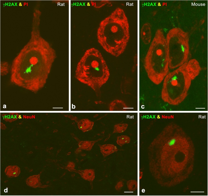
Fig. 1.

a-c Representative examples of immunolabeling for γH2AX of cortical neurons in a dissociated neuron preparation (a) and cryosections (b, c) counterstained with propidium iodide (PI) from irradiated rat (a, b) and mouse (c) at 15 days post-IR. Note the presence of typical PDDF associated with the nucleolus. d, e Rat cerebral cortex cryosections double immunolabeled for γH2AX and NeuN illustrate the specific localization of PDDF in NeuN-positive neurons at 15 days post-IR. Scale bars: a-c, e, 5 μm; d, 10 μm
