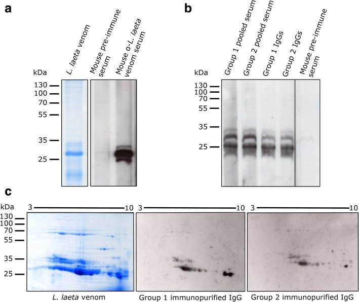
Fig. 2.
Immunoblot detection of L. laeta venom using pooled sera of Group 1 and Group 2. a Immunoblot detection of L. laeta venom with mouse anti-L. laeta venom immune serum. Lane 1: 12% SDS-PAGE of L. laeta venom stained with Coomassie brilliant blue. Lane 2: L. laeta venom immunoblot incubated with pre-immune mouse serum (1:1000 dilution). Lane 3: L. laeta venom immunoblot incubated with mouse L. laeta antivenom immune serum (1:10,000 dilution). b L. laeta venom immunoblot detected by pooled serum and purified IgGs of Group 1 or Group 2. Lanes 1 and 2: Serum pools for Group 1 and Group 2, respectively. Lanes 3 and 4: purified IgG antibodies (1 μg/mL) of Group 1 and Group 2 sera, respectively. Lane 5: pre-immune mouse serum. c Immunoblot of L. laeta venom separated by 2D electrophoresis

