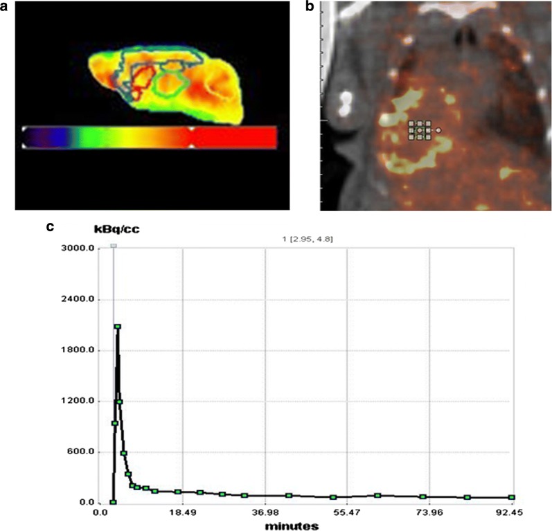
Fig. 1.
a Volume of interest (VOI) of mouse brain. VOI was drawn under guidance of the PMOD embedded mouse brain atlas (Mouse (M. Mirrione)-T2 MRI atlas) to cover the cortex (Cor: blue), right hippocampus (Rhip: dark green), left hippocampus (Lhip: dark green), thalamus (Thal: light green), right striatum (Rstr: red), left striatum (Lstr: red) and the cerebellum (Crbl: yellow), b volume of interest in blood input area, c time activity curve of blood input area

