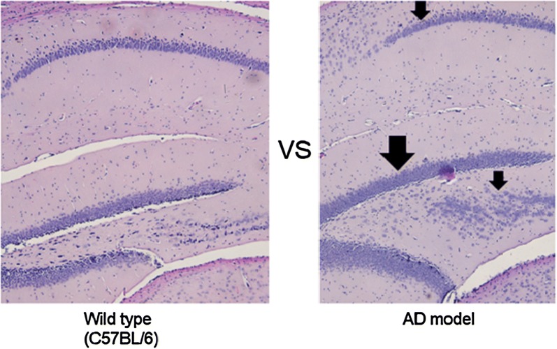
Fig. 7.
Visual comparison of the H & E staining image of hippocampus (sagittal section) between AD and wild type. Left: the zoom (100×) of the hippocampus in wild type, Right: the zoom (100×) of the hippocampus in AD mouse. AD mice show a more immature pattern as a result of disarrangement of hippocampal cell migration along the dentate gyrus of the hippocampus compared with the wild type

