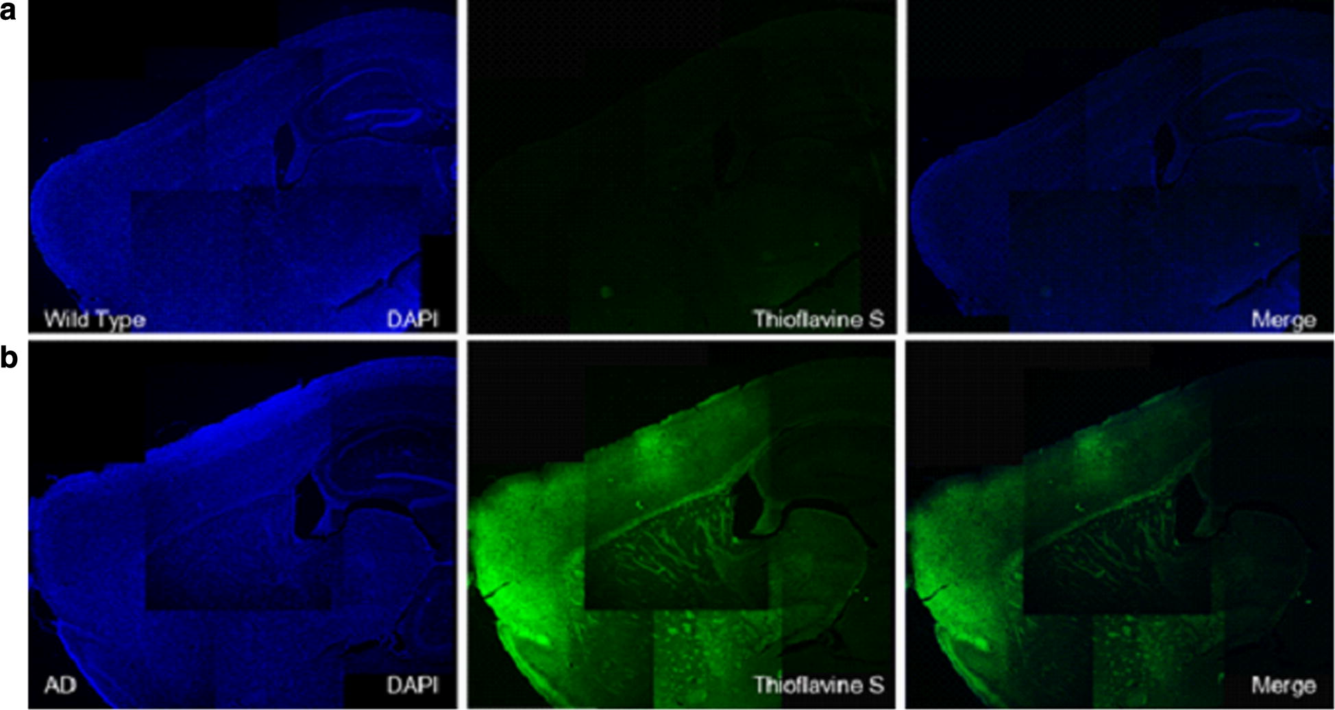
Fig. 8.

Visual overview of the thioflavin S staining images of a wild type mouse and b AD transgenic mouse. Left column shows DAPI (blue channel), middle column shows thioflavin S (green channel with specific staining signal) and right column shows merged image. Aβ deposits were found broadly in various brain regions including the cortex, hippocampus and thalamus in AD mice
