
Figure 6.
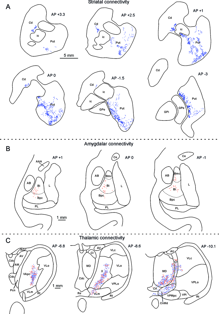
Striatal, amygdala and thalamus distribution of the labeling observed after Mk1 injection. (A). Drawings of coronal sections through the striatum showing the distribution of the anterograde labeling; the dot density is proportional to the density of the observed labeled terminals (one dot is equivalent to about 15–25 labeled terminals). The sections are shown in a rostral to caudal order and their AP level is indicated in terms of distance in mm from the anterior commissure (AC). Scale bar applies to all sections. Cd: caudate nucleus; GPe: external globus pallidus; GPi: internal globus pallidus; ic: internal capsule; Put: putamen. (B). Distribution of retrogradely labeled amygdalar neurons. Each dot corresponds to one labeled neuron. For each case, the labeling is shown in two drawings of coronal sections, selected at different AP levels. The dashed lines mark the borders of the magnocellular, intermediate, and parvocellular subdivisions of the basal nucleus. Scale bar applies to all sections. AAA: anterior amygdaloid area; AB: accessory basal nucleus; Bmc: magnocellular subdivision of the basal nucleus; Bi: intermediate subdivision of the basal nucleus; Bpc : parvocellular subdivision of the basal nucleus; Ce: central nucleus.L: lateral nucleus; PL: paralaminar nucleus. (C). Distribution of labeled thalamic neurons and terminals. The labeling is shown in drawings of coronal sections in rostral to caudal order selected at different AP levels according to the atlas of Olszewski (1952). Each red dot corresponds to a single labeled neuron, each blue dot is equivalent to about 15–25 labeled terminals. Scale bar applies to all sections. AM anterior medial nucleus, AV anterior ventral nucleus, Cl central lateral nucleus, Cn.Md centromedian nucleus, Csl central superior lateral nucleus, LD lateral dorsal nucleus, MD mediodorsal nucleus, MDmc mediodorsal nucleus, magnocellular part, MDmf mediodorsal nucleus, multiform part, MDpc mediodorsal nucleus, parvicellular part, Pcn paracentral nucleus, Pf parafascicular nucleus, Re reuniens nucleus, THI habenulointerpeduncular tract, TMT mammillothalamic tract, VAmc ventral anterior nucleus magnocellular part, VApc ventral anterior nucleus, parvicellular part, VLm ventral lateral nucleus, medial part, VLo ventral lateral nucleus, oral part, VPM ventral posterior medial nucleus, VPMpc ventral posterior medial nucleus, parvicellular part, X nucleus X of Olszewski (1952).